Joint Classification in Human Anatomy
Anatomy
Joints are the connection points between bones. Basically there are movable joints and immovable joints. For training purpose the first category is most interesting. Movable joints are divided into the highly mobile synovial joints, such as knee or shoulder joint, and cartilaginous joints, which allow only little movement, such as joints between sternum and ribs.
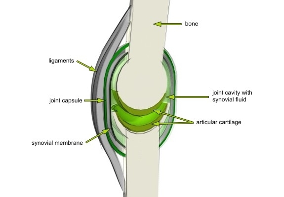
Synovial joints have a high range of motion because of a space between the articulating bones, which is filled with synovial fluid. The contact area of each bone is covered with a layer of cartilage, wich is elastically deformable, to protect it from pressure, friction and other forces.
Cartilage belongs to the supportive structures and has no artery. That´s why it is recovering or healing very slowly. It can only be nourished and supplied with oxygen by diffusion from surrounding tissues. The premise to that is multilateral movement to have the synoval fluids exchanged, waste products removed and the cartilage filled with nutrients.
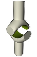
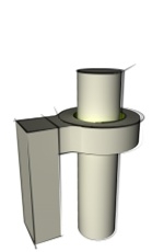
Synovial Joint Illustration
Synovial Joint Classification
| type | illustration | function | examples |
|---|
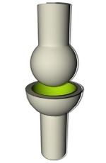
| Ball-and-Socket-Joint |
| Distal bone can move around a center in an indefinite number of axes. Main movements are flexion-extension, adduction-abduction, axial rotation and circumduction. | hips, shoulders |
|---|
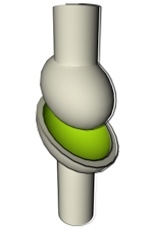
| Ellipsoid Joint |
| Distal bone has an ovoid articular surface and is received into an elliptical cavity, wich makes it impossible for the bones to do axial rotation. So main movements here are flexion-extension, adduction-abduction and circumduction. | wrist |
|---|
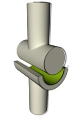
| Hinge Joint |
| Here the distal bone can move only in one plane, flexion and extension (forward and backward). | knee, elbow, fingers |
|---|
| Flat Joint |
| The main movements are flexion-extension and rotation. | wrist |
|---|
| Saddle Joint |
| The saddle joint consists of two opposing surfaces that are reciprocally concave-convex, which allows flexion, extension, adduction, abduction, and circumduction, but no axial rotation. | thumb |
|---|
| Pivot Joint |
| A pivot joint´s movement is limited to rotation. | Atlas and Axis, proximal radioulnar articulation |
|---|